Our blog
Resources and insights
The latest industry news, interviews, technologies, and resources.
Blog categories
Blog
P4 Allele of MYOZ3
IntroductionThis blog post explores the P4 allele (S42L) of the equine MYOZ3 gene, which encodes myozenin 3. Portions of this blog post serve as additional sources of information to supplement the MYOZ3 Gene Page.We present data to support the hypothesis that the P4 allele of MYOZ3 (S42L) is damaging.The substitution of leucine (hydrophobic) for serine (polar uncharged) in the P4 allele of MYOZ3 is a nonconservative substitution of a chemically dissimilar amino acid. Evolutionary conservation provides convincing evidence that the P4 allele of MYOZ3 is damaging. We use public data to show that the reference allele is completely conserved across 448 species of mammals and birds representing 155 unique sequences of the 41-amino acid query sequence centered on the S42L allele, covering over 310 million years of evolutionary history. P4 allele of equine MYOZ3Evolutionary conservation provides evidence on whether the P4 allele of MYOZ3 is damaging. In this approach, predicted MYOZ3 protein sequences are compared among a number of different species. This method, applied to mammals and birds, is shown below.A 41 amino acid segment of the protein sequence of equine MYOZ3 (XP_005599348.1), centered on the position of the variant, is shown below. This sequence is compared to a segment of the protein sequence of…
Blog
P2 Allele of MYOT
Introduction This blog post explores the P2 allele (S232P) of the equine MYOT gene, which encodes myotilin. Portions of this blog post serve as additional sources of information to supplement the MYOT Gene Page. We present data to support the hypothesis that the P2 allele of MYOT (S232P) is damaging. The substitution of proline (heterocyclic) for serine (polar uncharged) in the P2 variant of MYOT is a nonconservative substitution of a chemically dissimilar amino acid. Evolutionary conservation provides convincing evidence that the P2 allele of MYOT is damaging. We use public data to show that the reference allele is widely conserved across most of 244 species of mammals and birds covering over 310 million years of evolutionary history. There is a prominent exception in humans, where the MYOT gene has a conservative amino acid substitution that has gone to fixation (S232T). The substitution of threonine (polar uncharged) for serine (polar uncharged) in human MYOT is a conservative substitution of a chemically similar amino acid. This variant has gone to fixation in all the great apes most closely related to humans (chimp, bonobo, gorilla, and orangutan), but is absent from Old World monkeys, placing the origin in the common ancestor to…
Blog
Genes First!
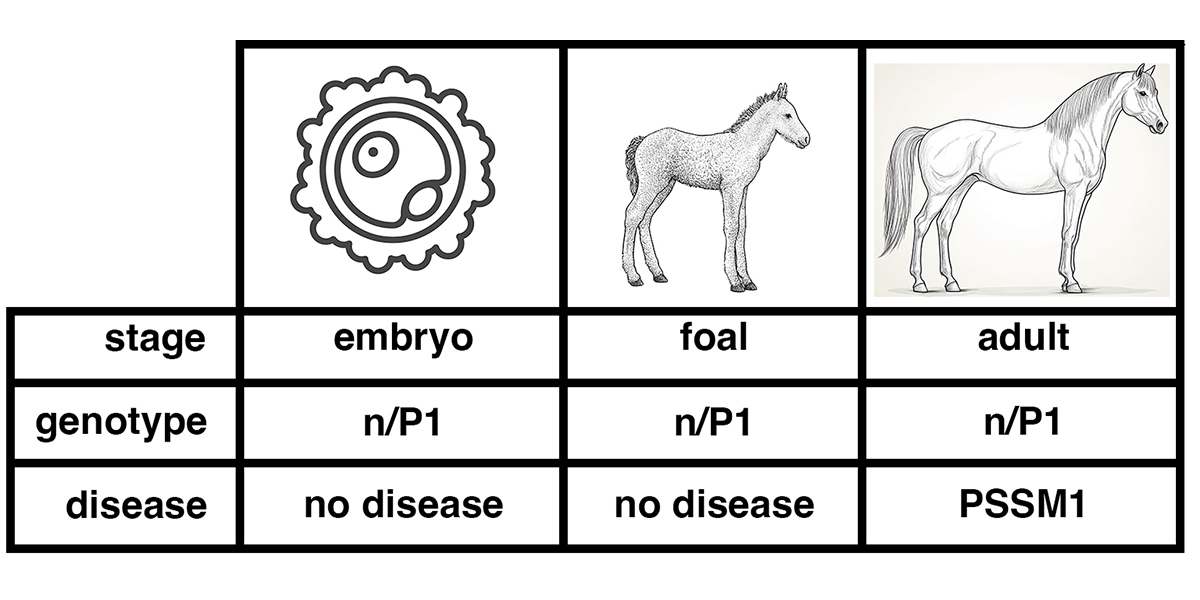
EquiSeq has released a major revision to the software that powers our website and database. We now present genetic test results starting with the affected gene. Here is an example of the appearance of the new horse profile pages. Figure 1. A horse profile page returning results from EquiSeq. This horse has tested positive for the K1 variant of COL6A3 (n/K1), negative for the other five variants in EquiSeq’s DNA test, and also negative for the P5 and P6 variants of DYSF, an experimental test not available commercially. What is a gene? In the context of mammalian genetics, a gene is a unit of hereditary information made of DNA. These segments of DNA encode proteins. Every mammal has two copies of every gene (except for X-linked genes in males). In a population, there are variant forms of genes, called alleles of that gene, that differ in their DNA sequence. Some alleles produce proteins that differ from the reference or wild-type sequence in minor ways that do not affect protein function. Other alleles produce proteins that are nonfunctional or have an altered function, causing a change in the development or function of the organism. What is a disease state? Disease is defined…
Blog
Study of horse genomes explores genetic burden
A team of researchers at the University of Minnesota and the University of California, Davis, have published a landmark study of the predicted genetic burden in horses, based on the analysis of whole genome sequence data from 605 horses (1). They conclude that the genetic load in horses is 1.4 – 2.6 times that of the human population. The authors discuss the unique advantages of the study of horse to understand human phenotypes, especially those associated with athletic performance.
Horse owners have asked us to explain this paper, as it mentions the genetic variants that are in EquiSeq’s panel of DNA tests. Here we review the methods and major findings of this paper. We include background information typically absent from the primary literature in order to make the paper more accessible to non-specialists.
Blog
What is PSSM?
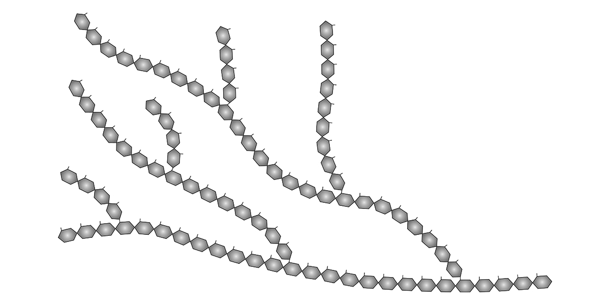
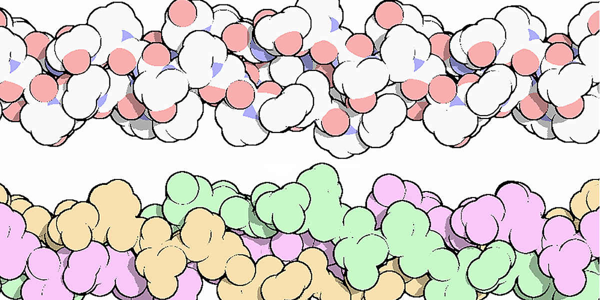
Polysaccharide Storage Myopathy (PSSM) is a form of equine exercise intolerance characterized by episodes of tying up. Horses with this condition have a particular set of clinical signs, including displays of pain, refusal to move forward, trouble standing for the farrier, and standing “parked out” as if to urinate. All of these are signs of a disease state affecting muscle. Affected horses often show enlarged granules of glycogen in muscle tissue obtained by biopsy. These enlarged glycogen granules are relatively resistant to digestion by amylase, an enzyme that completely degrades normal glycogen granules under standard conditions. Glycogen is a polysaccharide, a polymer of the simple sugar glucose, a monosaccharide. The synthesis of glycogen from glucose in muscle tissue provides a store of energy. When energy is needed, the glycogen is broken down into glucose, which can be metabolized for energy. The image above shows a) the chemical structure of glucose, b) the general structure of glycogen in which glucose units are joined in a branching structure, and c) the chemical structure of the linear polymers of glucose and the branch points. The synthesis of glycogen from glucose is highly regulated. In normal horses, glycogen is synthesized when energy needs are…
Blog
Genetic Linkage of P8 and K1
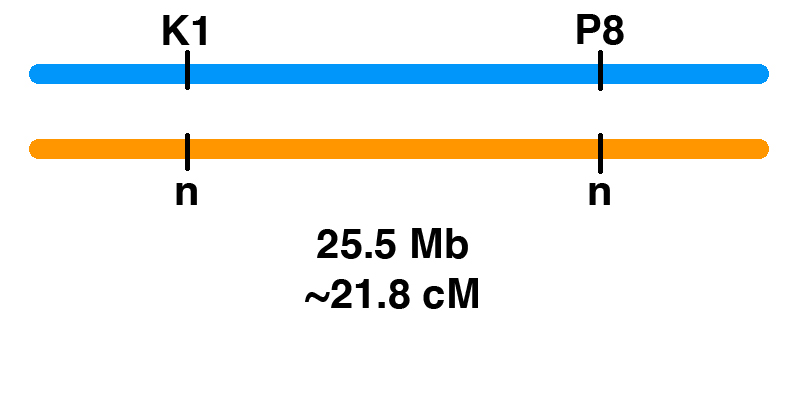
How can you figure out the chances of different genotypes when breeding horses? Monohybrid Cross It’s easy for one gene. Let’s say that there is a stallion that has one copy of one of the genetic variants associated with PSSM2, the P2 variant (MYOT-S232P). This stallion is heterozygous (n/P2), meaning that he has one normal copy of the MYOT gene and one copy with the P2 mutation. If the stallion is bred to a mare that is clear (n/n) for the P2 allele, the Punnett square below shows the odds. Gametes (sperm and eggs) contain a single copy of each gene in the horse. The stallion produces two kinds of sperm in equal frequency: sperm with P2, and sperm with the normal allele (n). The mare produces eggs with one of the two normal alleles (n). This means that there is a 50% chance of a foal that is n/P2, and a 50% chance of a foal that is n/n. Dihybrid Cross What if we are following two genes? In general, two gene pairs will show independent assortment. This means that the segregation of one pair of alleles into gametes will not influence the segregation of another pair of alleles….
Blog
The K1 Genetic Variant Affects COL6A3, a Gene Encoding a Collagen
Contents Introduction The K1 genetic variant that has been part of EquiSeq’s Myopathy Panel since October 2019 is a missense allele of COL6A3, a gene encoding a collagen [1]. Collagens are a family of proteins that are the main structural components of the connective tissue. Collagens also guide bone formation and play an important role in many other tissues. They are the most abundant proteins in the body of mammals. There are thirty types of collagen, made up of collagen proteins encoded by 44 different genes. In humans, mutations in different collagen genes are responsible for a wide variety of inherited disorders. The phenotype produced by mutations in different collagens gives us an insight into the role of each of these collagens. The COL6A3 gene encodes one of the type VI collagens that are components of the endomysium, a layer of connective tissue that covers individual muscle fibers. Mutations in human COL6A1, COL6A2, and COL6A3 are associated with Bethlem myopathy and Ullrich congenital muscular dystrophy [1-11]. The K1 allele of equine COL6A3 is defined by its coordinates in the current genome assembly for horse, chr6:23,416,882 C/G in EquCab3.0 [12]. For purposes of this blog post, we will use the protein model XP_023498413.1, which would make this allele G2178A, the substitution…
Blog Uncategorized
The P8 Genetic Variant Affects PYROXD1, a Gene Required for Oxidative Defense
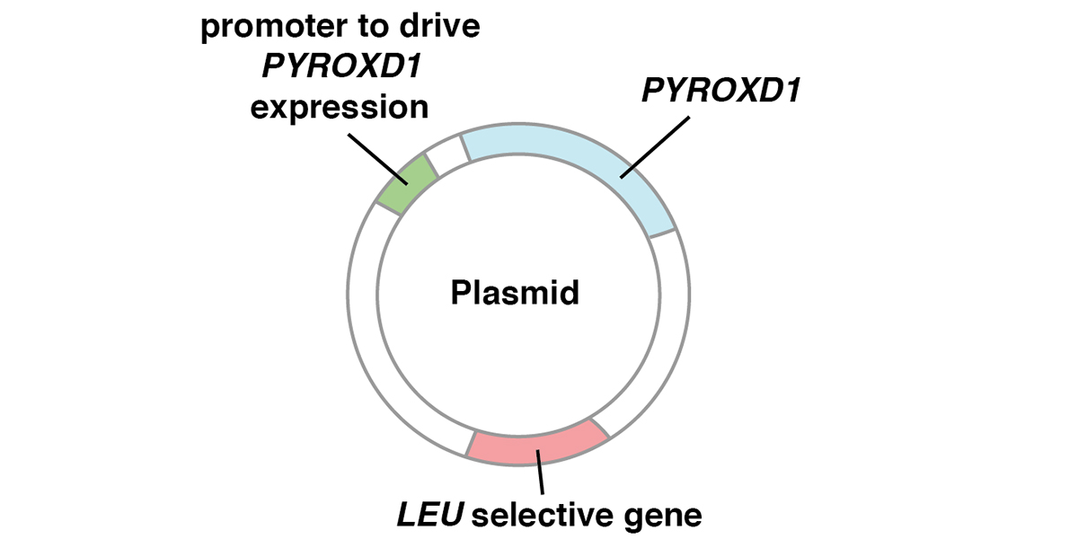
Contents Introduction The P8 genetic variant that has been part of EquiSeq’s Myopathy Panel since October 2019 is a missense allele of PYROXD1, a gene required for oxidative defense [1]. Mutations of the human PYROXD1 gene are associated with Myofibrillar Myopathy 8 in humans [1]. The P8 allele of equine PYROXD1 changes an aspartic acid (D) to a histidine (H), a nonconservative substitution. Comparison of the amino acid sequence of the PYROXD1 protein across 248 species of vertebrates shows that the position affected by the P8 allele of PYROXD1 is highly conserved across hundreds of millions of years of evolution, with only aspartic acid (D) or asparagine (N) seen in this position. Preliminary biochemical evidence suggests that the P8 allele of PYROXD1 reduces the stability of the protein. There is independent evidence that the basis of Arabian MFM is a defect in oxidative defense affecting the redox state of cysteine thiols. Function of PYROXD1 PYROXD1 encodes pyridine nucleotide-disulfide oxidoreductase domain 1, an FAD-binding oxidoreductase that uses NAD/NADH as a cofactor to reduce cysteine thiols as part of the cellular oxidative defense. Mutations in the human PYROXD1 gene are associated with Myofibrillar Myopathy 8 (MFM8), a disease state defined by a breakdown of the integrity of the sarcomeric Z disc and myofibrils, accompanied by…
Blog
Peer Review Fails to Identify Errors in Inconclusive Study of Genetic Variants of Equine Muscle Genes
This blog post is a discussion of a paper published by Williams et al. in a recent issue of Equine Veterinary Journal [1]. The paper aims to identify genetic variants that might be responsible for Myofibrillar Myopathy (MFM) in Warmbloods. The authors use a candidate gene approach, searching for variants of eight of the nine genes known to be associated with MFM in human patients and eight additional genes. Variants are identified by RNA-Seq of 16 warmbloods typed by muscle biopsy (8 affected and 8 control), and by analysis of additional whole genome sequence data available to the authors and in public databases. The citation is: Zoe J. Williams (will3084@msu.edu), Deborah Velez-Irizarry (no contact information), Jessica L. Petersen (jessica.petersen@unl.edu), Julien Ochala (julien.ochala@kcl.ac.uk), Carrie J. Finno (cjfinno@ucdavis.edu) and Stephanie J. Valberg (valbergs@cvm.msu.edu) (2020) Candidate gene expression and coding sequence variants in Warmblood horses with myofibrillar myopathy. Equine Vet J. 00: 1-10. PMID: 32453872. Contents Summary Ninety variants were identified in exons of thirteen of the sixteen genes (ACTA1, BAG3, DES, DNAJB6, FLNC, HSPB8, KY, LDB3, LMNA, MYOT, PLEC, PYROXD1, and SQSTM1). No coding variants were found for CRYAB, FHL1, or TIA1. Of the 90 variants, 62 were synonymous substitutions, 2 were splice region variants, and 26 were missense alleles. All 26 missense alleles (in 11 genes) were predicted to have moderate effects…
Blog
Genetic Basis of Exercise Intolerance in Arabians
Paul Szauter, PhD, Chief Scientific Officer of EquiSeq, gave a presentation titled “Genetic Basis of Exercise Intolerance in Arabians” at the Al Khamsa Annual Meeting and Convention in Fayetteville, Arkansas on October 13, 2019. EquiSeq is a company based in Albuquerque, New Mexico, that develops and sells genetic tests for horses. Exercise intolerance is known in Arabians (1, 2) and limits the performance of some affected horses in endurance events. Symptoms of exercise intolerance (tying up) include sudden lameness, myoglobinuria, and greatly increased serum levels of creatine kinase (CK), a muscle enzyme released into the bloodstream when muscles are damaged. Researchers from EquiSeq began studying exercise intolerance in Arabians at an endurance event at the Biltmore estate in 2017. They were stationed at the vet trailer in order to study horses pulled from the event for symptoms of lameness. The weather was unusually cold and rainy, and it wasn’t long before the first horse showed up at the vet trailer with urine the color of black coffee and obvious symptoms of pain and lameness. A test for serum CK (normally in the range of 500 or less) showed levels in excess of 20,000. Eight hours and thirty liters of IV…
